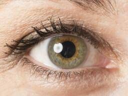
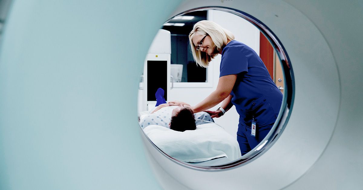
Optical coherence tomography, often shortened to OCT, sounds like something a spaceship might use to inspect an alien crystal. In real life, it is even more useful: it is a fast, noninvasive imaging test that lets doctors see tiny layers of living tissue in remarkable detail. Most people meet OCT at the eye doctor’s office, where it helps examine the retina, macula, optic nerve, and other delicate structures at the back of the eye. But this technology has also stepped beyond ophthalmology and into cardiology, dermatology, dentistry, neurology research, and other medical fields.
The easiest way to understand OCT is to think of it as an “optical ultrasound.” Ultrasound uses sound waves to create pictures of structures inside the body. OCT uses light waves instead. Because light has a much shorter wavelength than sound, OCT can capture extremely fine detailsespecially in transparent or semi-transparent tissues such as the eye. In practical terms, it allows clinicians to see microscopic layers that would otherwise stay hidden, like a medical version of lifting the lid on a lasagna and inspecting every noodle.
This article explains how optical coherence tomography works, what it is used for, what patients can expect during an OCT scan, and why this technology has become one of the most important diagnostic tools in modern eye care.
What Is Optical Coherence Tomography?
Optical coherence tomography is a medical imaging technique that creates high-resolution, cross-sectional images of tissue. In eye care, OCT is commonly used to scan the retina, the light-sensitive layer at the back of the eye. The scan can show the retina’s layers, measure their thickness, detect swelling, reveal fluid pockets, and help doctors monitor changes over time.
Unlike X-rays or CT scans, OCT does not use ionizing radiation. It relies on light, usually near-infrared light, which is directed toward the tissue being examined. The machine analyzes how that light reflects back from different depths. Those reflections are then converted into detailed images.
In ophthalmology, OCT has become a standard tool because many eye diseases begin with subtle structural changes long before a person notices blurry vision, distortion, or vision loss. A regular eye exam can tell a doctor a lot, but OCT adds a layer-by-layer view. It is like switching from a street map to satellite viewsuddenly, the hidden details start waving hello.
How Optical Coherence Tomography Works
OCT works through a principle called low-coherence interferometry. That phrase may sound like it escaped from a physics textbook, so let’s translate it into plain English.
The OCT device sends a beam of light toward the tissue. At the same time, part of the light travels along a reference path inside the machine. When the light reflected from the tissue returns, the system compares it with the reference light. By measuring tiny differences in timing and intensity, the machine determines how deep each reflection came from.
From Light Reflections to Medical Images
A single depth scan is called an A-scan. When many A-scans are combined side by side, they form a cross-sectional image called a B-scan. When many B-scans are collected across an area, the system can create a three-dimensional map of the tissue.
In retinal imaging, this means OCT can show the macula, optic nerve head, retinal nerve fiber layer, photoreceptor layer, and other important structures. Doctors can measure thickness, identify abnormal spaces, and compare today’s scan with previous scans. That comparison is one of OCT’s biggest strengths because slow changes can be easier to catch when the images are lined up over time.
Why Light Makes OCT So Detailed
Because OCT uses light rather than sound, it can achieve very high resolution. This is especially valuable in the eye, where the structures are tiny, layered, and transparent enough for light-based imaging. The retina is only a fraction of a millimeter thick, yet it contains multiple layers that play different roles in vision. OCT can separate those layers visually, giving doctors a detailed look at tissue architecture.
The result is not just a pretty picture. OCT provides measurable data. For example, in glaucoma care, doctors often evaluate the retinal nerve fiber layer and optic nerve structure. In macular disease, they may look for fluid, thinning, thickening, or disruption of outer retinal layers. The picture is useful, but the measurements are where the magic gets its clipboard.
What Is OCT Used For?
OCT is best known for eye care, but its usefulness comes from a broader idea: it can show fine tissue structure without cutting into the body. That makes it valuable wherever doctors need detailed surface or near-surface imaging.
OCT in Eye Exams
In ophthalmology and optometry, OCT is commonly used to diagnose and monitor conditions affecting the retina and optic nerve. It is especially helpful because many serious eye diseases can develop quietly. A patient may feel that their vision is “fine,” while OCT reveals early changes that deserve attention.
Common eye-related uses include:
- Age-related macular degeneration: OCT can help detect fluid, retinal thinning, and changes under or within the macula.
- Diabetic retinopathy and diabetic macular edema: OCT can identify swelling and fluid in the retina, which may affect central vision.
- Glaucoma: OCT can measure the retinal nerve fiber layer and optic nerve head, helping clinicians monitor structural damage.
- Macular hole: OCT can show the size, depth, and shape of a macular hole, helping guide treatment planning.
- Epiretinal membrane: OCT can reveal wrinkling or traction on the retinal surface.
- Retinal vein occlusion: OCT helps assess swelling and fluid after blocked retinal veins.
- Central serous chorioretinopathy: OCT can show fluid collecting under the retina.
For patients receiving eye injections, laser treatment, or surgery, OCT is often used to track whether the treatment is working. If fluid decreases, swelling improves, or the retina becomes more stable, the scan can help confirm progress.
OCT Angiography: Seeing Blood Flow Without Dye
OCT angiography, or OCTA, is a newer extension of OCT. Instead of showing only structure, OCTA detects motion from blood cells moving through vessels. This allows doctors to visualize the tiny blood vessels of the retina and choroid without injecting dye.
Traditional fluorescein angiography can still be very useful, especially when doctors need to see leakage patterns. However, OCTA offers a fast, noninvasive way to examine retinal and choroidal microvasculature. It may be used in conditions such as diabetic retinopathy, macular degeneration, retinal vascular disease, and glaucoma research.
OCTA is powerful, but it is not perfect. It can be affected by motion artifacts, poor fixation, media opacity, and segmentation errors. In other words, the machine is impressive, but it still appreciates a cooperative eyeball.
OCT in Cardiology
In cardiology, intravascular OCT can be used inside blood vessels to examine artery walls and coronary plaques. A tiny imaging catheter is placed inside the vessel, allowing doctors to see details that can help evaluate plaque characteristics, stent placement, and vessel healing. This is different from the eye scan version, but the core idea is the same: light-based imaging creates high-resolution pictures of tissue structure.
OCT in Dermatology
Dermatology has also adopted OCT in selected settings. Skin OCT can help visualize layers beneath the surface without immediately performing a biopsy. It has been studied for evaluating certain skin lesions, including basal cell carcinoma and other superficial skin conditions. While biopsy remains essential for many diagnoses, OCT can sometimes provide extra information before treatment decisions are made.
OCT in Dentistry and Other Fields
Researchers have explored OCT in dentistry for detecting early enamel changes, cracks, and restoration margins. OCT has also been studied in gastroenterology, neurology, pathology, and surgical guidance. The technology keeps expanding because many medical fields share the same wish: “Could we see this tiny structure clearly without making a bigger problem?” OCT often raises its hand.
What Happens During an OCT Scan?
An OCT eye scan is usually quick, painless, and contact-free. The patient sits in front of the machine, places their chin on a rest, and looks at a target light. The device scans the eye in seconds. Depending on the clinic, the whole process may take only a few minutes.
Sometimes the pupils are dilated with eye drops before the scan, especially if the doctor needs a clearer view of the retina. Dilation can make the eyes sensitive to light and may blur near vision for a few hours. Sunglasses become your temporary best friend, which is not a bad deal if you enjoy looking mysteriously important in a parking lot.
During the scan, the patient should try to stay still and blink when instructed. Dry eyes, cataracts, small pupils, or trouble focusing can reduce image quality. If the first scan is not clear, the technician may repeat it. That does not mean something is wrong; it often just means the machine wants a better pose from your retina.
Is Optical Coherence Tomography Safe?
For standard eye imaging, OCT is considered safe and noninvasive. It does not touch the eye and does not use radiation like an X-ray. The light exposure is carefully controlled by device standards. Most people do not feel anything during the scan.
However, OCT is a diagnostic tool, not a standalone answer machine. The scan must be interpreted by a trained clinician, and results need to be considered alongside symptoms, vision testing, eye pressure, medical history, and other exam findings. A beautiful OCT image can still be misleading if taken out of context. Medicine, unfortunately, has not yet installed a giant “obvious answer” button.
Benefits of OCT
OCT has become so widely used because it offers several practical advantages.
It Is Noninvasive
For routine eye OCT, there are no needles, no incisions, and no physical contact with the eye. This makes it comfortable for most patients and easy to repeat over time.
It Provides High-Resolution Detail
OCT can show tissue layers with impressive clarity. In retinal care, this can reveal swelling, fluid, holes, membranes, thinning, and structural changes that might not be obvious during a standard exam.
It Helps Track Disease Over Time
One scan is useful. A series of scans is even better. OCT allows clinicians to compare changes month by month or year by year. This is especially important in chronic conditions such as glaucoma, diabetic eye disease, and macular degeneration.
It Supports Treatment Decisions
OCT can help doctors decide whether treatment is needed, whether current treatment is working, or whether the treatment plan should change. For example, in wet macular degeneration, OCT can help monitor retinal fluid after therapy.
Limitations of OCT
OCT is excellent, but it is not a medical crystal ball. Several factors can limit its usefulness.
First, image quality depends on clear optical media. Cataracts, corneal scars, vitreous hemorrhage, or poor fixation may make scans less reliable. Second, OCT shows structure better than function. A retina may look fairly stable on OCT while vision is affected by another issue. Third, some findings require confirmation with other tests, such as visual field testing, fundus photography, fluorescein angiography, or a full clinical exam.
OCT also creates a lot of data. Automated measurements are helpful, but they can sometimes be thrown off by segmentation errors, unusual anatomy, or artifacts. That is why experienced interpretation matters. The machine measures; the clinician thinks. Ideally, both do their jobs and nobody gets dramatic.
Who Might Need an OCT Scan?
A doctor may recommend OCT if a patient has symptoms such as blurred central vision, distorted lines, unexplained vision loss, flashes or floaters, or signs of retinal disease. OCT may also be recommended for people with diabetes, glaucoma risk, age-related macular degeneration, optic nerve concerns, high myopia, retinal swelling, or a family history of certain eye conditions.
In many clinics, OCT is also used as part of routine monitoring for patients already diagnosed with eye disease. Because it is quick and repeatable, it helps clinicians detect small changes before they become big problems. That early warning ability is one reason OCT is sometimes described as one of the most important advances in modern ophthalmic imaging.
How OCT Results Are Interpreted
OCT results usually include images and numerical measurements. In a retina scan, the doctor may look at the fovea, macular thickness, retinal layers, and any signs of fluid or traction. In glaucoma evaluation, the scan may include optic nerve measurements, retinal nerve fiber layer thickness, and ganglion cell analysis.
Many OCT reports use color-coded maps, but patients should be careful not to interpret those colors alone. Green does not always mean “perfect,” and red does not always mean “panic immediately.” These colors compare measurements with a reference database, but normal anatomy varies. A trained clinician must interpret the findings in context.
The Future of Optical Coherence Tomography
OCT continues to evolve quickly. Faster scanners, wider fields of view, better depth imaging, adaptive optics, artificial intelligence, and home-based monitoring systems are all shaping the future of the technology. OCT angiography is expanding the ability to study microvascular disease. Intraoperative OCT is helping surgeons see tissue during delicate procedures. AI tools may help detect patterns, flag progression, and support clinicians who are reviewing large volumes of imaging data.
Still, the goal is not to replace doctors with machines. The goal is to give clinicians sharper tools and patients earlier answers. OCT is most valuable when it supports careful medical judgment, not when it tries to become the boss of the clinic.
Practical Experiences With Optical Coherence Tomography
For many patients, the first OCT scan is surprisingly simple. They may arrive expecting something intense because the name sounds complicated. Then they sit down, place their chin on the chin rest, look at a blinking target, and hear the technician say, “Great, we’re done.” The whole thing can feel almost suspiciously easy, like ordering a coffee and accidentally receiving a medical-grade map of your retina.
A common patient experience is curiosity. The OCT image often looks like a black-and-white landscape with hills, valleys, layers, and shadows. When a doctor points to fluid under the retina, thinning near the optic nerve, or a wrinkle on the macula, the abstract idea of an eye condition becomes more concrete. Patients can literally see the problem. That visual explanation can make treatment decisions easier to understand.
People being monitored for macular degeneration often become familiar with OCT over time. One visit may show fluid, another may show improvement after treatment, and another may help decide whether the next treatment interval can be adjusted. In this setting, OCT becomes less like a one-time test and more like a progress diary. The retina writes the entries; the machine translates them.
Patients with diabetes may also benefit from seeing OCT results. Diabetic macular edema can cause swelling in the central retina, and OCT can show whether fluid is present even when symptoms are mild. This can be a powerful reminder that eye health is closely linked to overall health. Blood sugar, blood pressure, cholesterol, and regular exams are not just numbers in a chart; they can affect the tissue responsible for sharp central vision.
For glaucoma patients, the experience is slightly different. Glaucoma often affects peripheral vision first, and many people do not notice early damage. OCT helps by measuring structures around the optic nerve and retinal nerve fiber layer. A patient may not feel any change, but OCT can help reveal whether structural thinning is stable or progressing. Combined with eye pressure checks and visual field tests, it gives the doctor a more complete picture.
From the clinic’s perspective, OCT improves communication. Instead of saying, “There may be swelling,” a clinician can show the scan and explain where the swelling is located. Instead of relying only on a patient’s memory of symptoms, the doctor can compare today’s scan with last year’s scan. This is especially useful because human memory is not always a precision instrument. Most of us can forget where we placed our keys within seven minutes; asking us to remember subtle vision changes over six months is ambitious.
Technicians also play a key role in the OCT experience. A good technician helps the patient sit comfortably, align correctly, blink at the right time, and stay relaxed. Small things matter. A dry eye, a blink, or a tiny movement can create artifacts. When the technician repeats a scan, it is often about quality control, not bad news.
The best experience with OCT happens when patients understand what the test can and cannot do. OCT can reveal structural details with amazing precision, but it does not replace a full eye exam. It does not measure every aspect of vision, and it does not automatically diagnose every disease. It is one part of the clinical story. When used properly, though, it is one of the clearest windows doctors have into living tissueespecially the retina.
Conclusion
Optical coherence tomography is a fast, noninvasive, light-based imaging technology that has transformed the way doctors examine the eye and other tissues. By using reflected light to create cross-sectional images, OCT allows clinicians to see microscopic layers, measure tissue thickness, detect fluid, monitor disease progression, and guide treatment decisions.
Its biggest impact has been in ophthalmology, where it helps diagnose and manage conditions such as macular degeneration, diabetic retinopathy, glaucoma, macular holes, and retinal swelling. Newer forms such as OCT angiography add vascular detail without dye injection, while research continues to expand OCT into cardiology, dermatology, dentistry, surgery, and artificial intelligence-assisted diagnostics.
For patients, OCT is usually quick, painless, and surprisingly low-drama. For doctors, it is a powerful tool that turns hidden tissue layers into visible, measurable information. In the world of medical imaging, OCT is proof that sometimes the brightest ideas really do involve light.