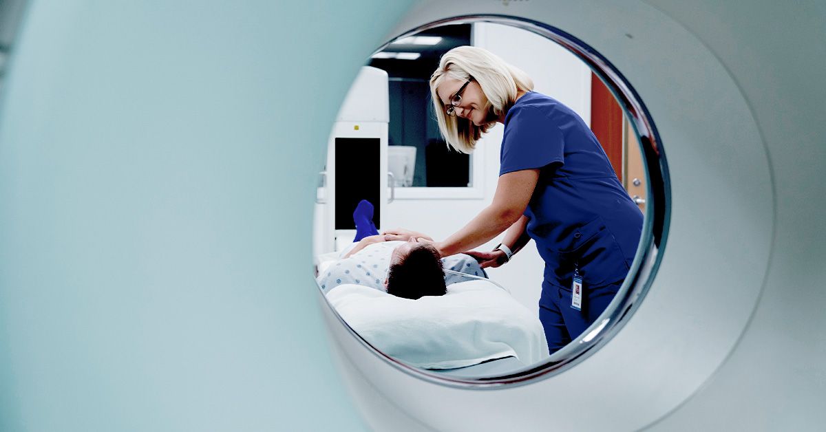
An MRI scan may sound like something borrowed from a science-fiction spaceship, but it is one of the most common and useful tools in modern medicine. MRI stands for magnetic resonance imaging. It helps doctors see detailed pictures of organs, tissues, joints, blood vessels, the brain, the spine, and other internal structures without using X-rays. In other words, it gives the body a high-resolution photo shoot while you lie still and try not to wonder why the machine sounds like a tiny construction crew with rhythm problems.
MRI scans are especially valuable when doctors need a closer look at soft tissues. While X-rays are excellent for bones and CT scans are often fast and useful in emergencies, MRI is famous for its ability to show fine detail in the brain, muscles, ligaments, spinal cord, organs, and many tumors. It can help diagnose disease, guide treatment, monitor healing, and sometimes answer the medical question everyone wants solved: “What exactly is going on in there?”
What Is an MRI Scan?
An MRI scan is a noninvasive imaging test that uses a powerful magnet, radio waves, and a computer to create detailed images of the inside of the body. “Noninvasive” means the scan itself does not involve cutting the skin or placing instruments inside the body. Most MRI exams are painless, although they can be noisy, lengthy, and a little uncomfortable if you dislike tight spaces.
Unlike X-rays and CT scans, MRI does not use ionizing radiation. Instead, the scanner’s magnetic field temporarily affects the behavior of hydrogen atoms in the body. Radio waves are then used to collect signals from those atoms. A computer turns those signals into images that radiologists can examine. The result is a detailed view of body structures, often sliced visually into thin sections, almost like looking through a medical loaf of breadless delicious, much more useful.
How MRI Scans Work
The human body contains a lot of water, and water contains hydrogen atoms. MRI technology takes advantage of this. When a person enters the MRI scanner, the machine’s strong magnetic field aligns many of these hydrogen atoms. Short bursts of radiofrequency energy then disturb that alignment. As the atoms return to their usual state, they release signals.
Different tissues release signals in different ways. Fat, muscle, fluid, scar tissue, inflammation, tumors, blood vessels, and organs can appear with different brightness or contrast on MRI images. That is why MRI can be so helpful for conditions where small tissue differences matter. The radiologist studies these images and sends a report to the doctor who ordered the test.
Common Uses of MRI Scans
Doctors order MRI scans for many reasons. The exact purpose depends on the body part being examined and the symptoms or medical history involved. MRI may be used to diagnose a new condition, evaluate an injury, plan surgery, check how well treatment is working, or monitor a known disease over time.
Brain and Nervous System
Brain MRI is often used to evaluate headaches with concerning features, seizures, dizziness, vision changes, hearing problems, stroke-like symptoms, tumors, multiple sclerosis, head injuries, and unexplained neurological symptoms. MRI can show the brain, spinal cord, nerves, and surrounding tissues in fine detail.
Spine and Back Problems
MRI is commonly used for back pain when doctors suspect a herniated disc, spinal stenosis, nerve compression, infection, tumor, or spinal cord problem. It can show discs, nerves, ligaments, and the spinal canal more clearly than a standard X-ray.
Joints, Bones, and Soft Tissues
Orthopedic specialists often use MRI to evaluate sports injuries and joint problems. It can help detect torn ligaments, cartilage damage, tendon injuries, bone infections, stress fractures, and soft tissue masses. Knee, shoulder, hip, ankle, and wrist MRI scans are especially common.
Heart and Blood Vessels
Cardiac MRI can help evaluate heart structure, heart muscle damage, congenital heart disease, blood flow, and certain vascular conditions. Magnetic resonance angiography, or MRA, may be used to look at blood vessels without the same type of radiation exposure used in some other imaging tests.
Abdomen and Pelvis
MRI can help examine the liver, kidneys, pancreas, uterus, ovaries, prostate, bladder, bowel, and other abdominal or pelvic structures. It may be used to evaluate tumors, inflammation, pelvic pain, liver disease, prostate concerns, uterine fibroids, endometriosis, and other conditions.
Cancer Diagnosis and Monitoring
MRI is often part of cancer care. It may help detect tumors, define their size and location, guide biopsy or treatment planning, and monitor response after chemotherapy, radiation, surgery, or other therapy. Breast MRI, prostate MRI, brain MRI, and liver MRI are examples where MRI may provide important detail.
Types of MRI Scans
Not every MRI is the same. The scan is customized based on what the doctor needs to see. A brain MRI focuses on the head and nervous system. A spine MRI examines the neck, mid-back, or lower back. A musculoskeletal MRI studies joints, muscles, tendons, and ligaments. A cardiac MRI looks at the heart. A breast MRI is often used for high-risk screening or cancer evaluation. A functional MRI, or fMRI, measures changes in blood flow related to brain activity.
Some MRI exams are done without contrast. Others use contrast material to make certain tissues, blood vessels, inflammation, or tumors easier to see. Whether contrast is needed depends on the medical question being asked.
MRI With Contrast: What It Means
MRI contrast is a special substance that may be injected into a vein during the exam. The most common MRI contrast agents contain gadolinium. Contrast can make certain areas appear clearer on the images, helping doctors identify inflammation, tumors, infection, blood vessel abnormalities, or scar tissue.
Most people tolerate MRI contrast well. Possible side effects may include temporary nausea, headache, dizziness, a cool feeling during injection, or mild discomfort at the IV site. Allergic reactions are uncommon, but they can happen. People with serious kidney disease need special screening because gadolinium-based contrast agents can rarely cause serious complications in patients whose kidneys cannot clear the substance properly.
If you are pregnant, breastfeeding, have kidney disease, have had a previous reaction to contrast, or have concerns about contrast material, tell your healthcare team before the scan. The goal is not to scare anyone; it is to make sure the MRI is done safely and for the right reason.
How to Prepare for an MRI Scan
Preparation depends on the type of MRI. For many routine MRI scans, you can eat, drink, and take your regular medications as usual. Some abdominal, pelvic, or sedation-related exams may require fasting or special instructions. Always follow the directions from the imaging center or your healthcare provider.
Before the Appointment
You may be asked detailed safety questions before your MRI. This screening is important because the MRI magnet is powerful and always deserves respect. Tell the staff if you have a pacemaker, defibrillator, cochlear implant, aneurysm clip, implanted pump, neurostimulator, artificial heart valve, metal fragments, surgical clips, shrapnel, or any other implanted medical device.
Some newer medical devices are MRI-safe or MRI-conditional, meaning they may be allowed under specific conditions. Others are not safe in the MRI environment. Do not guess. Bring device information cards if you have them, and let the MRI staff verify everything.
What to Wear
You will usually change into a gown or approved clothing. Leave jewelry, watches, hairpins, removable dental work, hearing aids, wallets, credit cards, phones, keys, and body piercings outside the scan room. Some clothing contains metallic fibers, snaps, zippers, or antimicrobial metals, so even your favorite yoga pants may be politely rejected at the MRI door. The machine is not judging your style; it is protecting your skin.
Claustrophobia and Anxiety
If tight spaces make you anxious, tell your doctor or imaging center ahead of time. Options may include relaxation techniques, music, eye coverings, a support person nearby when allowed, an open MRI scanner, or medication for anxiety. If sedation is planned, you may need someone to drive you home.
What Happens During the MRI Procedure?
When it is time for the scan, a technologist helps position you on a padded table. The body part being scanned may be placed inside a special device called a coil, which helps receive signals and improve image quality. The table then slides into the MRI scanner, which is shaped like a large tube in many facilities.
During the scan, the technologist watches from another room and communicates with you through an intercom. You will usually be given earplugs or headphones because MRI machines make loud knocking, tapping, buzzing, and thumping sounds. Think of it as industrial music with a medical degree.
The most important job during the scan is to stay still. Movement can blur the images, and blurry images may require repeat sequences. Depending on the exam, you may be asked to hold your breath for short periods. If contrast is needed, the technologist may place an IV before or during the scan.
A typical MRI can take about 15 minutes to more than an hour, depending on the body part, the number of images needed, whether contrast is used, and whether extra sequences are required. The scan itself should not hurt. You may feel warmth in the area being scanned, which can be normal, but you should report burning, pain, shortness of breath, itching, or sudden discomfort immediately.
After the MRI Scan
After a routine MRI without sedation, most people can return to normal activities right away. If contrast was used, your body usually clears it through urine. Drinking fluids may be recommended unless your doctor has told you to limit fluids.
If you received sedation or anti-anxiety medication, you may feel drowsy and need someone to take you home. Follow the discharge instructions given by the imaging center.
When Do You Get MRI Results?
MRI images are interpreted by a radiologist, a doctor trained in medical imaging. The radiologist prepares a report and sends it to the healthcare provider who ordered the test. Your doctor then explains the results in the context of your symptoms, exam, medical history, and other tests.
Timing varies. Some results are available quickly, especially in urgent situations. Routine outpatient results may take a few days. Seeing the images before the explanation can be confusing, so avoid diagnosing yourself from a patient portal at midnight. The internet is wide, dramatic, and not always invited to medical decision-making.
Is an MRI Safe?
MRI is considered safe for many people when proper screening and safety procedures are followed. The magnetic field itself is not known to harm the average person, and MRI does not expose patients to ionizing radiation. However, MRI safety depends heavily on identifying metal objects, implants, devices, kidney problems, pregnancy status, and prior contrast reactions.
The strongest safety rule is simple: be honest and thorough during screening. Even small details matter. A forgotten implant, metal fragment, medication patch, or device can change the plan. MRI teams ask many questions not because they enjoy paperwork, but because magnets are extremely serious about their job.
Who May Not Be Able to Have an MRI?
Some people may not be candidates for MRI, or they may need special arrangements. This can include patients with certain pacemakers, implanted defibrillators, older aneurysm clips, cochlear implants, metal fragments near the eye, some implanted pumps, or devices that are not approved for MRI. People with severe kidney disease may need extra caution before receiving gadolinium contrast.
Pregnancy does not automatically rule out MRI, but the decision should be made carefully with a healthcare provider. Gadolinium contrast is generally avoided during pregnancy unless the information is essential and cannot be obtained another way.
MRI vs. CT Scan vs. X-Ray
MRI, CT scans, and X-rays are all imaging tools, but they are not interchangeable. X-rays are fast and often used for bones, chest imaging, and dental exams. CT scans use X-rays and computer processing to create detailed cross-sectional images; they are fast and often used in emergencies, trauma, lung disease, and many abdominal problems. MRI takes longer but provides excellent soft tissue detail and does not use ionizing radiation.
The “best” test depends on the medical question. A suspected broken wrist may need an X-ray first. A major trauma case may need a CT scan. A possible ligament tear, brain tumor, spinal cord issue, or multiple sclerosis evaluation may need MRI. Good imaging is not about choosing the fanciest machine; it is about choosing the right tool.
Benefits of MRI Scans
MRI offers several major benefits. It provides detailed images of soft tissues, helps doctors detect problems that may not appear clearly on other tests, and can be repeated when medically necessary without radiation exposure. It can also reduce the need for exploratory procedures in some situations by giving doctors a clearer internal map.
MRI can be especially useful for complex diagnoses. For example, a person with persistent knee pain after a sports injury may have a normal X-ray but an MRI can reveal a torn meniscus. A patient with neurological symptoms may need a brain MRI to look for inflammation, tumors, stroke changes, or demyelinating disease. A person undergoing cancer treatment may receive MRI scans to see whether a tumor is shrinking, stable, or growing.
Limitations and Possible Downsides
MRI is powerful, but it is not perfect. It can be expensive, may take longer than other imaging tests, and can be uncomfortable for people with claustrophobia. Some patients cannot have MRI because of implanted devices or metal. MRI can also find incidental findingsunexpected abnormalities that may or may not matter. These can lead to helpful early diagnosis, but sometimes they cause extra testing and anxiety.
Another limitation is that MRI requires stillness. Young children, people with severe pain, and patients who cannot remain still may need sedation or a different imaging method. The scanner is also noisy, and the enclosed design can be stressful. Still, for many medical questions, the information gained is worth the temporary inconvenience.
How to Make an MRI More Comfortable
A little preparation can make the experience easier. Ask how long the scan may take. Use the restroom beforehand. Wear comfortable clothing that is easy to change out of. Leave valuables at home. Practice slow breathing before the exam. If music is offered, choose it. If you are nervous, tell the technologist. MRI staff help anxious patients every day; you are not the first person to treat the scanner like a suspicious tunnel.
During the scan, focus on small goals. Stay still for one sequence at a time. Count breaths. Imagine a calm place. Remind yourself that the noises are normal. The technologist can see and hear you, and most scanners provide a call button. You are not locked in, and you can communicate if something feels wrong.
Patient Experience: What an MRI Really Feels Like
For many people, the hardest part of an MRI is not painit is anticipation. You may arrive worried about the results, nervous about the machine, or annoyed that you had to remove every metal object you own, including the watch you forgot was on your wrist. The first few minutes can feel strange because the room is controlled, the equipment looks serious, and everyone is carefully following safety steps.
Once you are on the table, the technologist positions you carefully. This can feel surprisingly specific. A knee MRI may involve cushions and supports around the leg. A brain MRI may include a frame-like coil around the head. A shoulder MRI may require your arm to rest in a certain position. The goal is not to make you feel like a museum exhibit; it is to keep the area still and centered so the images are sharp.
When the table moves into the scanner, people react differently. Some feel perfectly calm. Others feel a wave of “Oh, absolutely not.” If you are prone to claustrophobia, closing your eyes before the table moves can help. Many patients prefer not to look around once inside. The scanner is well lit and ventilated, but the space can still feel close.
Then come the sounds. MRI noise is not subtle. You may hear tapping, knocking, buzzing, humming, and rhythmic thumping. The noises change as the machine collects different image sequences. Ear protection helps, and some centers offer headphones with music. The sound does not mean something is wrong. It means the scanner is working, even if it sounds like a dishwasher arguing with a printer.
Staying still is the main challenge. At first, it seems easy. Then your nose itches, your foot wants to twitch, or your brain suddenly remembers a grocery list from 2019. Try to relax your muscles and breathe normally unless told otherwise. If you move, the technologist may need to repeat part of the scan, which means more time in the machine. Stillness is the fastest exit strategy.
If contrast is used, you may feel a small IV pinch. Some people notice a cool sensation during injection. Most feel nothing dramatic. Tell the team immediately if you feel itching, trouble breathing, swelling, dizziness, or anything unusual. Serious reactions are rare, but MRI teams are trained to respond.
After the scan, many patients feel relief simply because it is over. The exam may have been noisy, boring, awkward, or mildly stressfulbut usually not painful. The bigger emotional moment often comes later, while waiting for results. That waiting period can be difficult. A helpful approach is to remember that MRI is a tool for clarity. It does not create the problem; it helps reveal what may need attention, treatment, monitoring, or reassurance.
Conclusion
MRI scans are one of medicine’s most valuable imaging tools. They use magnets, radio waves, and computer technology to create detailed pictures inside the body without ionizing radiation. Doctors use MRI to evaluate the brain, spine, joints, organs, heart, blood vessels, tumors, injuries, inflammation, and many other conditions.
The procedure is usually painless, but it requires preparation, safety screening, stillness, and patience. Contrast may be used when doctors need extra detail, and some patients need special precautions because of kidney disease, pregnancy, implanted devices, or metal in the body. If your doctor recommends an MRI, ask what question the scan is meant to answer, how to prepare, whether contrast is needed, and when you can expect results.
The machine may be loud, the tunnel may feel dramatic, and the gown may not win any fashion awards. But when used appropriately, MRI can give doctors a remarkably clear view of what is happening inside the bodyand that clarity can make a real difference in diagnosis, treatment, and peace of mind.
Note: This article is for general educational purposes only and does not replace medical advice, diagnosis, or treatment from a qualified healthcare professional.