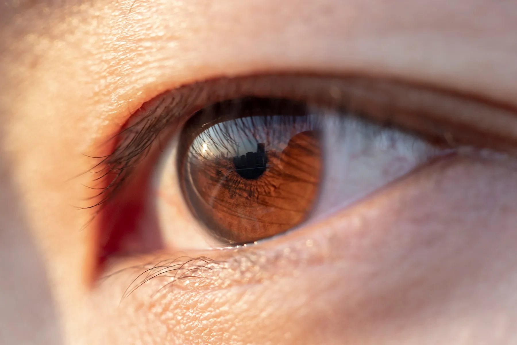
When people hear that diabetes can affect the eyes, they often imagine needing stronger glasses or squinting harder at restaurant menus. Unfortunately, diabetic eye disease is not usually impressed by a new prescription. It can damage the retina, the delicate tissue at the back of the eye that turns light into signals the brain can understand.
One of the most important complications is diabetic macular edema, commonly shortened to DME. It develops when damaged retinal blood vessels leak fluid into the maculathe small but extremely hardworking area responsible for sharp central vision. As fluid accumulates, the macula swells, and everyday tasks such as reading, driving, recognizing faces, or working on a computer may become difficult.
DME can be serious, but a diagnosis does not automatically mean blindness. Modern retinal imaging can detect subtle swelling, and treatments can reduce fluid, stabilize vision, and sometimes improve sight. The key is catching the problem before permanent retinal damage has had time to unpack its bags and settle in.
What Is Diabetic Macular Edema, Exactly?
Diabetic macular edema is swelling in the macula caused by diabetes-related damage to tiny blood vessels in the retina. These weakened vessels may allow blood components, proteins, and fluid to escape into nearby retinal tissue.
The retina is a thin layer of light-sensitive tissue lining the back of the eye. Near its center is the macula, which supplies the detailed, straight-ahead vision needed to:
- Read words and numbers
- Recognize faces
- See fine details
- Drive safely
- Use phones and computers
- Distinguish colors clearly
At the center of the macula is the fovea, a tiny area that provides the sharpest vision. When swelling involves this central zone, doctors may call the condition center-involved diabetic macular edema. Even a small amount of fluid in such a visually important location can make the world appear as though someone smeared petroleum jelly across the center of a camera lens.
How Is DME Related to Diabetic Retinopathy?
Diabetic macular edema and diabetic retinopathy are closely connected, but they are not interchangeable terms.
Diabetic retinopathy is the broader disease in which chronically elevated blood glucose damages retinal blood vessels. Early-stage disease is called nonproliferative diabetic retinopathy. In more advanced proliferative diabetic retinopathy, the retina begins growing fragile, abnormal blood vessels that may bleed or produce scar tissue.
Diabetic macular edema is a particular complication involving fluid accumulation and swelling in the macula. It usually occurs in someone who already has signs of diabetic retinopathy, but it can develop during different stages of that disease. In other words, retinopathy is the larger plumbing problem; DME is what happens when the leak reaches the eye’s high-definition viewing area.
What Causes Diabetic Macular Edema?
Blood vessels normally keep fluid and other substances inside the bloodstream. Long-term exposure to high blood glucose can damage the cells that maintain this protective barrier. Retinal capillaries may weaken, become blocked, bulge into tiny microaneurysms, or start leaking.
Damaged or oxygen-deprived retinal tissue can also produce increased amounts of vascular endothelial growth factor, or VEGF. This protein promotes blood-vessel growth and increases vascular leakage. In DME, too much VEGF can contribute to continued fluid accumulation inside the retinal layers.
Inflammatory processes may play a role as well. That helps explain why anti-VEGF medicines work well for many people, while some patients benefit from treatments that target inflammation through corticosteroids. DME is therefore not one simple leak with one universal repair kit. Its biology and response to treatment can vary from eye to eye.
Who Is at Risk?
Anyone with type 1 or type 2 diabetes can develop diabetic eye disease. Risk generally increases with the duration of diabetes, especially when blood glucose, blood pressure, or cholesterol remains above an individual’s target range.
Important risk factors include:
- Living with diabetes for many years
- Blood glucose levels that are frequently above target
- High blood pressure
- Abnormal cholesterol or triglyceride levels
- Existing diabetic retinopathy
- Kidney disease related to diabetes
- Pregnancy in someone with preexisting diabetes
- Smoking or other factors that damage blood vessels
- Missing recommended dilated eye examinations
Good diabetes management can substantially reduce risk, but it cannot make anyone completely invincible. A person with apparently well-managed diabetes may still develop retinal changes, particularly after many years. That is why “My eyesight seems fine” is not a substitute for an examination. Early diabetic retinopathy and DME may be present before obvious symptoms appear.
Symptoms of Diabetic Macular Edema
DME is usually painless. You cannot feel the retina swelling, and the condition may initially affect only one eye or progress more quickly in one eye than the other. Because the clearer eye can compensate, changes may go unnoticed until both eyes are affected or the swelling becomes more pronounced.
Possible symptoms include:
- Blurred or hazy central vision
- Straight lines appearing bent or wavy
- Words looking distorted while reading
- Colors appearing faded or washed out
- Difficulty recognizing faces
- Problems seeing fine details
- Dark, blank, or smudged areas near the center of vision
- Vision that seems to fluctuate from one day to another
- Increasing difficulty driving, especially when reading signs
Blurred vision does not always mean DME. Temporary blood glucose changes can affect the eye’s focusing ability, and cataracts, glaucoma, dry eye, medication effects, and other retinal conditions can also alter vision. Only an eye examination can identify the actual cause.
When should you seek urgent care?
Contact an eye-care professional promptly for new or worsening central blur, distortion, or color changes. Sudden flashes of light, a rapid increase in floaters, a curtain-like shadow, abrupt vision loss, severe eye pain, or significant redness may indicate another urgent eye problem and should be evaluated immediately.
How Doctors Diagnose DME
A routine vision test alone cannot rule out diabetic macular edema. Diagnosis usually requires a comprehensive dilated eye examination and retinal imaging.
Dilated retinal examination
Dilating drops widen the pupils so the doctor can inspect the retina and optic nerve. The examination may reveal retinal thickening, leaking microaneurysms, small hemorrhages, fatty deposits known as hard exudates, or other signs of diabetic retinopathy.
Optical coherence tomography
Optical coherence tomography, or OCT, is one of the most useful tests for DME. It uses light waves to create detailed cross-sectional images of the retina. OCT can measure retinal thickness, show pockets of fluid, determine whether the fovea is involved, and help doctors compare the eye’s response from one appointment to the next.
Retinal photography
Color or wide-field photographs document the condition of the retina. These images provide a visual record that can reveal changes over time.
Fluorescein angiography
In selected cases, a dye is injected into a vein, and a camera tracks it as it passes through retinal blood vessels. Fluorescein angiography can identify areas of leakage, poor circulation, and abnormal vessel growth. It may be especially useful when the diagnosis is uncertain or laser treatment is being considered.
How Is Diabetic Macular Edema Treated?
Treatment depends on the location and severity of swelling, the amount of vision loss, the condition of the other eye, previous treatments, and the patient’s overall health. Some people need prompt injections, while others may be monitored closely at first.
1. Anti-VEGF injections
Anti-VEGF therapy is a major treatment for center-involved DME, particularly when vision has declined. The medicine is injected into the vitreous, the gel-like substance inside the eye, where it blocks VEGF and helps reduce leakage and swelling.
The words “injection into the eye” understandably cause many patients to consider moving to a remote island with no forwarding address. In practice, the eye is numbed and cleaned beforehand, and the needle is extremely small. Patients may notice pressure, mild irritation, or temporary floaters, but the procedure is generally brief.
Treatment often begins with a series of closely spaced injections. The schedule may later be adjusted according to vision and OCT results. Some eyes improve substantially; others mainly stabilize, and a portion respond incompletely. Keeping appointments matters because long treatment gaps can allow swelling and vision loss to return.
2. Corticosteroid treatment
Steroid injections or longer-lasting steroid implants may reduce inflammation and vascular leakage. They are sometimes considered when anti-VEGF therapy has not produced an adequate response, when frequent visits are difficult, or when individual eye characteristics make steroids appropriate.
Steroids can raise eye pressure and increase the likelihood of cataracts, so patients require careful monitoring. They are useful tools, but not the sort that should be borrowed casually from the neighbor’s garage.
3. Laser treatment
Focal or grid laser treatment was once the primary therapy for DME. Anti-VEGF medicines are now commonly favored for center-involved disease with vision loss, but laser remains useful in selected cases, particularly when leakage is located away from the foveal center.
The laser applies controlled energy to leaking areas of the retina. Its main goal is generally to reduce continued leakage and lower the risk of further visual decline rather than instantly restoring perfect vision.
4. Vitrectomy surgery
Surgery is not needed for most cases of DME. A vitrectomy may be recommended when scar tissue or the vitreous is pulling on the macula, when there is persistent bleeding, or when another structural retinal problem is contributing to vision loss.
5. Observation with close follow-up
Not every visible pocket of retinal fluid demands immediate treatment. If DME involves the center of the macula but visual acuity remains good, a retina specialist may recommend close observation in appropriate cases. Treatment can begin if vision worsens or imaging shows concerning progression. Observation is still an active medical plannot permission to disappear until the next presidential election.
Managing Diabetes Is Part of Eye Treatment
Eye injections treat what is happening inside the retina, but they do not eliminate the underlying vascular effects of diabetes. Long-term management usually involves cooperation among the patient, ophthalmologist or retina specialist, primary-care clinician, endocrinologist, and sometimes a kidney or cardiovascular specialist.
Important goals may include:
- Keeping blood glucose within the individualized target range
- Managing blood pressure
- Treating unhealthy cholesterol levels
- Taking prescribed medications consistently
- Avoiding tobacco
- Choosing a nutrient-dense eating pattern
- Remaining physically active when medically appropriate
- Managing kidney and cardiovascular conditions
Rapid improvement in blood glucose can occasionally be associated with short-term worsening of existing retinopathy in certain circumstances. That does not mean someone should keep glucose high. It means major treatment changes should be coordinated with medical and eye-care professionals, particularly when advanced retinopathy is already present.
Can Diabetic Macular Edema Be Prevented?
No prevention strategy offers a 100% guarantee, but the risk of DME and severe vision loss can often be reduced. The most effective approach combines diabetes management with regular retinal screening.
People with diabetes should follow their clinician’s recommended schedule for comprehensive dilated eye examinations. Depending on the type of diabetes, pregnancy status, previous findings, and disease severity, examinations may be needed annually or more frequently.
Do not wait for blurred vision. The retina is annoyingly polite in the early stagesit may suffer quietly without sending a complaint. Detecting leakage or retinopathy before noticeable sight loss creates more opportunities to protect vision.
Living With DME
DME can affect independence, employment, driving, reading, medication management, and emotional well-being. Practical adaptations may help while treatment is underway:
- Increase text size and contrast on digital devices.
- Use brighter, directed lighting for reading and cooking.
- Keep medications in clearly labeled or talking organizers.
- Ask about low-vision rehabilitation if visual limitations persist.
- Arrange transportation for dilated examinations or procedures.
- Bring a written medication list and glucose records to appointments.
- Report visual changes instead of waiting for the next scheduled visit.
It is also reasonable to feel anxious, frustrated, or tired of medical appointments. DME care may require repeated monitoring and treatment. Asking the retina team what the OCT shows, what improvement is expected, and what would trigger a change in therapy can make the process feel less mysterious.
Frequently Asked Questions
Can DME cause blindness?
Yes. Untreated or advanced DME can cause severe and sometimes permanent central vision loss. It usually does not erase all peripheral vision, but central damage can make reading, driving, and facial recognition extremely difficult. Early detection and treatment significantly improve the chance of preserving useful sight.
Can diabetic macular edema go away?
Swelling may resolve or improve with treatment and better management of systemic health. However, DME can recur, persist, or require long-term therapy. A dry-looking retina on one OCT scan does not necessarily mean the condition is permanently cured.
Will glasses fix DME?
Glasses can correct ordinary focusing errors, but they cannot remove fluid from the retina. A new prescription may help after the macula has stabilized, but retinal swelling itself requires monitoring or medical treatment.
Is treatment painful?
Most patients experience pressure or brief discomfort rather than sharp pain during an injection because numbing medicine is used. Mild irritation afterward is common. Increasing pain, redness, light sensitivity, discharge, or rapidly worsening vision after an injection requires immediate contact with the treating clinic.
Does DME affect both eyes?
It can affect one or both eyes. Even when both are involved, the severity may differ. Each eye is evaluated and treated according to its own visual acuity, imaging findings, and treatment response.
A Realistic DME Experience: From “Dirty Glasses” to a Treatment Plan
The following is a fictional composite based on experiences commonly reported by patients. It is not the story of a specific individual and should not be interpreted as medical advice.
Imagine a 58-year-old office manager named Daniel who has lived with type 2 diabetes for 14 years. His days involve spreadsheets, invoices, email, and the mysterious office printer that jams only when someone is in a hurry.
One Monday morning, Daniel notices that the figures in the center of his computer screen look slightly fuzzy. He cleans his glasses. The blur remains. He cleans the monitor, adjusts the brightness, enlarges the text, and briefly suspects that the accounting software has launched a personal attack.
Over the next several weeks, he struggles to read street signs from the usual distance. Faces look clear around the edges but soft in the middle. When he covers his left eye, the lines on a window frame appear slightly bent through his right eye. Because there is no pain, redness, or dramatic loss of sight, he assumes he needs new glasses.
At an optometry appointment, the doctor sees signs of diabetic retinopathy and refers Daniel to a retina specialist. Dilating drops make the waiting-room lights look as bright as a movie premiere, but the examination itself is painless. Retinal photographs show small hemorrhages and deposits. An OCT scan reveals fluid in the center of the right macula and milder swelling in the left eye.
The retina specialist explains that Daniel has center-involved diabetic macular edema. Hearing the word “edema” is manageable. Hearing that treatment may involve an eye injection is considerably less charming. Daniel asks whether there is a pill, a stronger pair of glasses, an herbal tea, or perhaps a firm handshake that could solve the problem instead.
The specialist explains why anti-VEGF treatment is recommended for the right eye and why the left eye can be monitored closely for now. Daniel agrees to the injection after the staff describes each step. His eye is numbed, disinfected, and held open with a small device. He feels pressure for a moment. The anticipation turns out to be worse than the procedure.
That evening, the eye feels mildly gritty. A temporary spot floats through his vision, but it gradually disappears. Daniel follows the clinic’s instructions and knows which warning signs require an urgent call.
He receives additional injections over the following months. Progress is not cinematic. There is no orchestra, no instant crystal-clear sunrise, and no moment when he reads microscopic print from across the room. Instead, OCT images show the fluid slowly decreasing. His vision becomes more stable, and reading spreadsheets requires less magnification.
Meanwhile, Daniel meets with his primary-care clinician. They review his glucose records, blood pressure, kidney tests, cholesterol, medications, eating habits, and activity level. He begins taking short walks after meals, prepares lunches more often, and sets phone reminders for medication. These changes do not replace retinal treatment, but they support his broader vascular health.
Daniel also learns to prepare for appointments. He brings questions, arranges transportation when dilation will affect his driving, and asks the specialist to compare each OCT with the previous scan. Instead of focusing only on whether his vision is “better,” he starts understanding whether the retina is drier, stable, or worsening.
There are frustrating weeks. An appointment is delayed by work. One scan shows that a little fluid has returned. Daniel worries that treatment has failed, but the specialist explains that recurrence is common and adjusts the plan. DME management is often a long conversation rather than a single victorious speech.
After a year, Daniel’s right-eye vision has improved modestly, and the swelling is much better controlled. The left eye remains stable under observation. He still uses larger text for long reports, but he drives during daylight, recognizes faces, and continues working.
His biggest lesson is not that every patient will have the same outcome. They will not. It is that subtle blur deserves attention, treatment requires persistence, and an eye that does not hurt can still need urgent care. He also stops treating annual dilated examinations like optional software updates. Some reminders are genuinely worth clicking.
The Bottom Line
Diabetic macular edema is a diabetes-related buildup of fluid in the macula, the part of the retina responsible for sharp central vision. It may cause blur, distortion, faded colors, and difficulty reading or recognizing faces, although early disease can be symptom-free.
A dilated examination and OCT imaging can detect and measure the swelling. Depending on visual acuity and retinal findings, treatment may include anti-VEGF injections, corticosteroids, laser therapy, surgery, or careful observation. Managing blood glucose, blood pressure, cholesterol, kidney health, and other vascular risks remains an essential part of protecting sight.
Most importantly, do not wait for vision to become noticeably poor before arranging diabetic eye care. The sooner retinal changes are found, the more opportunities the medical team has to preserve the details that make daily life recognizable.