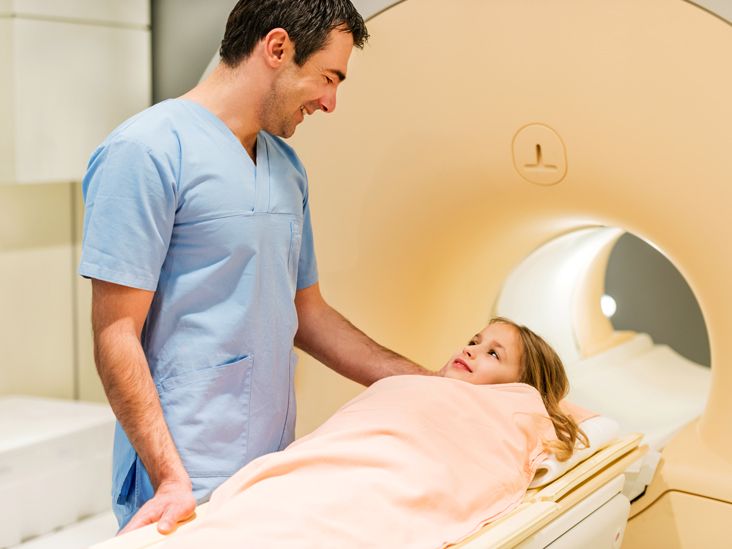
If you’ve ever wondered whether ADHD shows up on a brain scan like a broken bone on an X-ray, you’re not alone. Many people imagine a doctor pointing at a colorful image and saying, “See that red blob? That’s your ADHD.” Reality is… more complicated, less dramatic, and honestly a lot more interesting.
Brain scans do reveal important clues about attention-deficit/hyperactivity disorder (ADHD). Researchers using MRI, fMRI, PET, and SPECT imaging have spent decades studying how the brains of people with ADHD work compared with those without the condition. They’ve found real, measurable differences in brain structure and activity. But there’s a big catch: those differences are seen at the group level, not the individual “this-scan-equals-ADHD” level.
Major organizations like the American Academy of Pediatrics (AAP), the American Psychiatric Association (APA), the National Institute of Mental Health (NIMH), and the Centers for Disease Control and Prevention (CDC) all agree on one key point: brain imaging is not currently a stand-alone diagnostic test for ADHD. Instead, ADHD is diagnosed the old-fashioned waythrough careful history, behavior reports, and clinical evaluation.
So what can a brain scan reveal about ADHD? Let’s walk through what we know, what’s still emerging, and what it realistically means for you or your child.
ADHD and the Brain: A Quick Refresher
ADHD is a brain-based condition, not a character flaw
ADHD is classified as a neurodevelopmental disorder, meaning it involves differences in how the brain grows and functions over time. People with ADHD can experience persistent patterns of inattention, hyperactivity, and impulsivity that interfere with daily life at school, work, or home.
These symptoms are linked to brain circuits involved in attention, executive function (planning, organizing, prioritizing), motivation, emotional regulation, and reward. If you’ve ever felt like your brain’s “tab manager” is glitchy or your internal “brake pedal” responds a little too late, you’ve gotten a taste of how ADHD can feel from the inside.
Key brain regions involved in ADHD
Neuroimaging research has repeatedly pointed to several areas that tend to differ, on average, in people with ADHD:
- Prefrontal cortex: Involved in planning, decision-making, and self-control.
- Basal ganglia: Helps regulate movement, habit formation, and reward processing.
- Cerebellum: Important for coordination and timing, including timing of thoughts and actions.
- Networks connecting frontal areas to deeper brain regions that manage attention and inhibition.
Some studies have found slightly smaller volumes or delayed maturation in these regions in children with ADHD, with some “catching up” as they grow older. But again, these are subtle statistical trendsnot all-or-nothing differences you can reliably see in a single person’s scan.
Can ADHD Be Seen on a Brain Scan?
The short answer: not in a yes/no way
Researchers can use brain scans to compare large groups of people with and without ADHD. On average, they see patterns of structural and functional differences. But when you zoom down to an individual scan, the variability is huge. Plenty of people without ADHD have “ADHD-like” features on imaging, and many with ADHD have scans that look completely typical.
The authors of the DSM-5-TR (the manual clinicians use to diagnose mental health conditions) state clearly that no biological marker is diagnostic for ADHD, and that current meta-analyses of neuroimaging do not produce a fingerprint that reliably separates individuals with ADHD from those without it.
Types of brain scans you might hear about
- MRI (magnetic resonance imaging): Shows detailed brain structureuseful for ruling out tumors, malformations, or injury.
- fMRI (functional MRI): Measures changes in blood flow related to brain activity while someone performs tasks or rests.
- PET (positron emission tomography): Uses small amounts of radioactive tracers to measure metabolism or neurotransmitter activity.
- SPECT (single photon emission computed tomography): Another type of nuclear imaging that shows blood flow and activity patterns.
- EEG / QEEG: Measures electrical activity; sometimes marketed as “ADHD brain mapping.”
All of these technologies can show differences at the group level between people with ADHD and those withoutbut none of them act like a pregnancy test where one result means “ADHD” and the other means “no ADHD.” That’s why major professional bodies caution against using any of them as stand-alone diagnostic tools.
What Big Medical Organizations Say About ADHD Brain Scans
American Academy of Pediatrics (AAP)
The AAP’s clinical guidelines for diagnosing ADHD in children and teens focus on a thorough history, standardized symptom checklists, and reports from families and schoolsnot imaging tests. They do not recommend routine brain scans for ADHD diagnosis. Imaging may be used only when there are red flags suggesting another neurological problem that needs to be ruled out.
American Psychiatric Association (APA)
The APA has published position statements noting that current neuroimaging technologies do not yet offer clinically useful biomarkers for diagnosing or guiding treatment in most psychiatric conditions, including ADHD.
Centers for Disease Control and Prevention (CDC) and NIMH
CDC and NIMH emphasize that ADHD is diagnosed through a comprehensive evaluation of symptoms and functioning across settings. They highlight imaging as a valuable research tool to better understand ADHD, but not as a routine clinical test.
Bottom line: if a website or clinic promises to “diagnose ADHD definitively from one brain scan,” that’s more marketing than medicine.
What Brain Scans Can Reveal About ADHD
Clues about brain networks, not a single “ADHD spot”
Recent large-scale studies, including research analyzing more than 10,000 functional brain images of youth with ADHD, have found that ADHD is linked to atypical interactions between the frontal cortex and deeper information-processing regions. These findings support the idea that ADHD involves network-level differences in how attention, inhibition, and reward are coordinated.
Other work suggests that some children with ADHD show delayed cortical maturationessentially, parts of the brain may develop on a “different timetable” compared with peers, especially in regions handling attention and executive function.
Potential future uses: prognosis and treatment prediction
Researchers hope that, one day, imaging patterns might help predict which children are more likely to outgrow ADHD symptoms, who might respond best to certain medications or behavioral therapies, or who might be at risk for co-occurring conditions like anxiety or mood disorders. For now, these ideas are promising but still firmly in the research categorynot everyday clinical practice.
When a Brain Scan Actually Makes Sense in ADHD Care
Even though brain scans don’t diagnose ADHD, they can still be medically useful in certain situations. A clinician might order imaging if:
- There are unusual neurological signs (seizures, vision loss, weakness, severe headaches).
- The child or adult has a history of serious head trauma, stroke, or another brain condition.
- Symptoms started suddenly or worsened rapidly in a way that doesn’t fit a typical ADHD pattern.
In those cases, MRI or CT is used to look for other medical problemstumors, bleeding, structural abnormalitiesnot to “see ADHD” itself.
What about SPECT and other “functional” scans sold directly to patients?
Some clinics promote SPECT or other functional imaging as a way to “see” ADHD and tailor treatment. There is ongoing debate here. Some nuclear medicine groups have endorsed clinical uses of SPECT in psychiatry, and certain studies suggest SPECT patterns can differentiate adults with ADHD from controls.
However, major psychiatric associations still consider this evidence preliminary and do not endorse routine SPECT scanning to diagnose ADHD or dictate treatment. It’s important to know that SPECT and PET involve exposure to small doses of radiation, are expensive, and may not be covered by insurance when used for nonstandard indications.
Should You Get a Brain Scan for Suspected ADHD?
For most people, the answer is: probably not necessaryunless your doctor is worried about another neurological problem or there are red-flag symptoms. The standard of care for diagnosing ADHD is still a thorough clinical evaluation, which may include:
- Detailed history of symptoms across childhood and adulthood.
- Input from parents, teachers, partners, or coworkers.
- Standardized rating scales and checklists.
- Screening for other conditions like anxiety, learning differences, sleep issues, or depression.
Think of it this way: ADHD is diagnosed by how it affects your life, not by how it looks on a picture.
If you’re curious about brain scans because you want “proof” that ADHD is real, that’s completely understandable. It can be validating to see physical evidence. Just remember that a good clinician doesn’t need an MRI to believe youand neither should you.
Important note: This article is for general information only and can’t replace personalized medical advice. If you’re wondering whether imaging makes sense in your situation, talk directly with your health care provider.
How to Talk With Your Doctor About Brain Imaging and ADHD
If brain scans are on your mind (no pun intended), here are some questions you might ask your clinician:
- “Based on my symptoms and history, do you see any reason to order a brain scan?”
- “Are there red-flag signs that make you worried about something other than ADHD?”
- “What might a scan change about my treatment planif anything?”
- “Are there risks, including radiation or cost, that I should know about?”
A thoughtful clinician will explain whether imaging is medically indicated, and they should be open to discussing why they door don’trecommend it in your case.
Lived Experiences: What a Brain Scan Meant for People With ADHD
Research papers and policy statements are helpful, but it’s often the human stories that stick with us. Here are a few types of experiences people with ADHD describe when brain scans become part of their journey. These are composite examples based on common themesnot real individualsbut they reflect what many people report.
The person who wanted “proof”
Alex spent years hearing that they were “lazy” and “just not trying hard enough.” When they finally got an ADHD diagnosis as an adult, they still felt a nagging doubt: “What if I’m just making excuses?” After reading about brain scans online, Alex became convinced that an imaging test would finally validate their experience.
When Alex brought this up, their psychiatrist walked them through the evidence: brain scans show average differences for groups, not yes/no results for individual people. The doctor compared it to height chartsuseful for populations, less meaningful for one person standing alone. They focused instead on how clearly Alex’s symptoms matched ADHD criteria and how much those symptoms improved with treatment and coping strategies.
Alex never got a brain scan. Instead, they started tracking their progress in a journal. Over time, seeing concrete improvementsturning in work on time, remembering appointments, finishing tasksfelt more validating than any high-tech image could have.
The parent who feared something “worse” than ADHD
When Maya’s 8-year-old son, Leo, started having trouble focusing and sitting still, his teacher suggested an ADHD evaluation. But Maya was worried about something more seriouswhat if it was a brain tumor, epilepsy, or another scary neurological condition?
Leo’s pediatrician carefully reviewed his history, did a neurological exam, and asked about headaches, seizures, vision changes, and other red-flag symptoms. None were present. The doctor explained that in cases like Leo’swhere the pattern fits typical ADHD and there are no warning signsbrain imaging usually doesn’t add useful information.
Instead of ordering an MRI right away, the doctor followed evidence-based ADHD guidelines: behavior rating scales, input from school, and a stepwise treatment plan. As Leo responded well to behavioral strategies and, eventually, medication, Maya’s fear that something “worse” was lurking in his brain gradually eased.
The patient whose scan ruled out other problems
Jordan, a college student with long-standing ADHD, suddenly started having severe headaches and brief episodes of vision loss. These symptoms were new and worryingvery different from the usual distractibility and procrastination.
Jordan’s doctor didn’t assume it was “just the ADHD acting up.” Instead, they ordered an MRI to look for structural problems. The scan helped rule out tumors and other serious neurological issues. While the imaging didn’t show ADHD itself, it was crucial in making sure nothing more dangerous was going on.
Jordan still managed their ADHD with medication, therapy, and campus accommodationsbut they were grateful that the scan had answered the scariest question: “Is there something seriously wrong with my brain?”
The person drawn in by brain-scan marketing
Sam saw online ads promising personalized ADHD treatment based on “cutting-edge brain scans.” The idea sounded amazing: a customized plan built on a visual map of their brain function. The price, however, was in the thousands of dollars and would not be covered by insurance.
After reading up on the science and talking with a trusted clinician, Sam realized that while these scans might offer interesting information, they weren’t proven to lead to better outcomes than standard, guideline-based ADHD treatment. Sam ultimately chose to invest in evidence-based caremedication, therapy, coaching, and structured routinesrather than an expensive scan whose benefits were unclear.
Sam’s takeaway: “It’s cool that neuroscience is advancing, but I don’t need a glamorous brain picture to justify taking my ADHD seriously.”
What these stories have in common
Across all these experiences, one theme stands out: brain scans can sometimes play a helpful supporting roleespecially when ruling out other conditionsbut they’re not the main character in the ADHD story. The starring role still belongs to careful clinical evaluation, honest conversations, and practical strategies that help people function better in daily life.
If you’re curious about what a brain scan might show in ADHD, that curiosity is valid. Use it as a springboard for informed conversations with your health care teamnot as a reason to doubt yourself if no one orders a high-tech test.
Sources (for reference, not for display on page):
CDC ADHD overview and diagnosis information
American Academy of Pediatrics ADHD diagnosis guidelines
NIMH ADHD topic and research on brain structure and networks
DSM-5-TR and reviews on lack of diagnostic biomarkers for ADHD
Reviews and studies on ADHD neuroimaging, structural and functional differences
APA and related commentary on limits of neuroimaging in psychiatric diagnosis
AAN practice advisory on QEEG and ADHD
Research and opinion pieces on SPECT imaging and ADHD
Popular-press and educational overviews on ADHD brain scanning